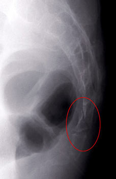

Maigne JY, Tamalet B (1996) Standardized radiologic protocol for the study of common coccygodynia and characteristics of the lesions observed in the sitting position. Kim NH, Suk KS (1999) Clinical and radiological differences between traumatic and idiopathic coccygodynia.
#Coccyx fracture series
Maigne JY, Rusakiewicz F, Diouf M (2012) Postpartum coccydynia: a case series study of 57 women. Hermel MB, Gershon-Cohen J (1953) The nutcracker fracture of the cuboid by indirect violence. Minutes of the 1st international symposium on coccyx pain, Maîtrise Orthopédique, pp 87–93īeckmann NM, Chinapuvvula NR (2017) Sacral fractures: classification and management. In: Maigne J-Y, Doursounian L (ed) Coccyx disorders. Woon JT, Perumal V, Maigne JY, Stringer MD (2013) CT morphology and morphometry of the normal adult coccyx. Lateral roentgenograms in the sitting position and coccygeal discography. Maigne JY, Guedj S, Straus C (1994) Idiopathic coccygodynia.

Tekin L, Yilmaz B, Alaca R, Ozçakar L, Dinçer K (2010) Coccyx fractures in patients with spinal cord injury. Raissaki MT, Williamson JB (1999) Fracture dislocation of the sacro-coccygeal joint: MRI evaluation. Hamoud K, Abbas J (2015) Fracture dislocation of the sacro-coccygeal joint in a 12-year-old boy. Kaushal R, Bhanot A, Luthra S, Gupta PN, Sharma RB (2005) Intrapartum coccygeal fracture, a cause for postpartum coccydynia: a case report. Gutierrez PR, Mas Martinez JJ, Arenas J (1998) Salter-Harris type I fracture of the sacro-coccygeal joint. Maigne JY, Doursounian L, Chatellier G (2000) Causes and mechanisms of common coccydynia: role of body mass index and coccygeal trauma. These slides can be retrieved under Electronic Supplementary Material. This should help the clinician in the management of these patients. Conclusionsįor the first time, a classification of fractures of the coccyx is presented. Although often thought to be one solid piece of bone, the coccyx can. Furthermore, due to the nature of the coccyx structure, many tailbone fractures are not immediately diagnosed. Nutcracker and obstetrical fractures were instable in their majority. A fractured coccyx can be a very painful acute or chronic ordeal which can be blindingly symptomatic and may not resolve for a very long duration. Flexion fractures usually healed spontaneously, but an associated intermittent luxation was possible. Flexion fractures (38 cases) involved the upper coccyx in 35 cases, and in 3 cases with a perineal trauma, it was the lower coccyx compression fractures (24 cases) involved the middle coccyx and occurred only when Co2 was square or cuneiform and Co3 was long and straight, hence a nutcracker mechanism four patients were adolescents with a compression of the sacrum extremity and were labeled adolescent compression fracture of S5 (type 2b) extension fractures (38 cases) were obstetrical and involved the lower coccyx their key feature was a progressive separation of the fragments with time. Three mechanisms are proposed to describe these fractures: flexion, compression and extension (types 1, 2 and 3, respectively). The mechanism, level, characteristics of the fracture line and complications were recorded. MethodsĪ series of 104 consecutive patients with a fracture of the coccyx was studied.

To describe a classification of fractures of the coccyx, according to their mechanism.


 0 kommentar(er)
